

In the field of medicine and cardiology, The full form of ECG stands for Electrocardiogram. An electrocardiogram is a diagnostic test that records the electrical activity of the heart over a period of time. It is a valuable tool for healthcare professionals to assess the heart’s health, detect irregularities, and diagnose various heart conditions. In this comprehensive guide, we will explore the details of ECG, its purpose, the procedure involved, and its significance in cardiology and patient care.
What is an ECG?
An Electrocardiogram (ECG or EKG, from the German word “Elektrokardiogramm”) is a non-invasive medical test that measures and records the electrical activity of the heart. It provides valuable information about the heart’s rhythm, rate, and the presence of any abnormalities or conditions affecting the heart’s function.
Purpose of an ECG
ECGs serve several essential purposes in cardiology and healthcare:
Diagnosis:
ECGs are used to diagnose a wide range of heart conditions, including arrhythmias, myocardial infarctions (heart attacks), and heart diseases.
Monitoring:
They are employed to monitor the heart’s electrical activity over time, especially for patients with chronic heart conditions or during surgery.
Assessment of Medications:
ECGs can assess the effects of medications on the heart and determine if they are working as intended.
Preoperative Evaluation:
Before surgery, ECGs help assess a patient’s cardiac health and determine their ability to tolerate surgery and anesthesia.
Screening:
In some cases, ECGs are used for heart disease screening, particularly for individuals at higher risk.
ECG Procedure
The ECG procedure involves the following steps:
- Electrode Placement: Small, adhesive electrodes are placed on specific areas of the patient’s chest, arms, and legs. These electrodes are connected to the ECG machine.
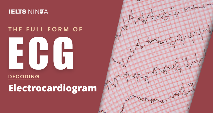
- Recording: The ECG machine records the electrical impulses generated by the heart as a series of waveforms on graph paper or a digital screen.
- Analysis: Healthcare professionals analyze the recorded ECG to assess the heart’s rhythm, rate, and any irregularities.
- Interpretation: The ECG results are interpreted to make a diagnosis or assess the heart’s condition.
Common ECG Waves and Intervals
ECG recordings typically include several waves and intervals, including:
- P-Wave: Represents atrial depolarization (contraction).
- QRS Complex: Depicts ventricular depolarization (contraction).
- T-Wave: Corresponds to ventricular repolarization (relaxation).
- PR Interval: Measures the time it takes for electrical impulses to travel from the atria to the ventricles.
- QT Interval: Measures the time it takes for ventricular depolarization and repolarization.
Also Read: Best online IELTS coaching & training academy
Significance of ECG in Cardiology
ECGs are of significant importance in cardiology for the following reasons:
Early Detection:
They can detect heart conditions at an early stage, allowing for timely intervention and treatment.
Risk Assessment:
ECGs help assess the risk of cardiac events in patients with known heart diseases.
Treatment Guidance:
They guide treatment decisions, such as medication choices and the need for procedures like cardiac catheterization.
Postoperative Care:
ECGs are used to monitor patients after cardiac surgery to detect complications.
Research:
ECG data is valuable for research into heart diseases and arrhythmias.
Conclusion
An Electrocardiogram (ECG) is a fundamental diagnostic tool in cardiology and healthcare that provides critical insights into the electrical activity of the heart. It is essential for diagnosing heart conditions, monitoring patients, guiding treatment decisions, and ultimately improving patient outcomes.